Research on swine dysentery (SD) and porcine proliferative enteropathy (PPE) requires an integrated understanding of pathogen localization, mucosal function, and the intestinal environment. Brachyspira primarily affects the mucus and mucosa of the cecum and colon, whereas Lawsonia intracellularis infects crypt epithelial cells in the ileum. Mucosal injury or proliferation can impair nutrient utilization, and coinfection may exacerbate disease. Intervention studies have observed reduced clinical SD with specific fiber combinations, inhibition of Brachyspira by commensal bacterial supernatants, and improvements in certain intestinal endpoints after administration of a complex fermented preparation in a defined Lawsonia inoculation model. These findings connect pathogen ecology, digestive function, and microbial materials while showing that growth, pathogen, and mucosal endpoints have distinct biological meanings. [1, 2, 3, 5, 6, 7, 8]
Pathogen Biology Determines the Primary Site of Lesions
Brachyspira hyodysenteriae is an anaerobic spirochete and a classical cause of SD. Strongly beta-hemolytic B. hampsonii and B. suanatina can produce similar disease; therefore, diagnostics should identify the appropriate species rather than treating every Brachyspira-positive result as swine dysentery. Intestinal spirochetosis caused by B. pilosicoli generally produces a milder form of colitis and should be differentiated from classical mucohemorrhagic SD. [1, 13]
Lawsonia intracellularis is a microaerophilic, obligate intracellular bacterium that mainly infects proliferating crypt epithelial cells in the terminal ileum; lesions may also extend into the jejunum or large intestine. PPE may be subclinical, present as chronic growth impairment, or manifest as an acute hemorrhagic condition. Pigs without obvious diarrhea may nevertheless experience reduced nutrient utilization and growth. Mucosal necrosis in SD and crypt proliferation in PPE are central diagnostic distinctions, although disease severity also depends on infection stage and host factors. [2, 11]
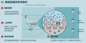
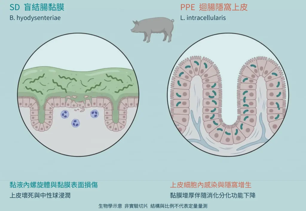 Figure 1. Colonic Mucosal Damage Versus Ileal Crypt Proliferation — Original biological concept diagram, not an experimental histological image; based on references [1, 2]. Left: Brachyspira-associated cecocolonic mucus and surface injury. Right: intracellular Lawsonia infection and proliferation of crypt epithelial cells. No experimental samples or statistical tests. Created by HYGEM using BioRender with Chinese annotations.
The luminal mucus environment shown on the left differs fundamentally from the intracellular infection target on the right. This distinction helps explain why inhibition by microbial supernatants, mucosal protection, and clinical disease outcomes should be measured separately.
Mucus Barrier and Inflammatory Changes in Swine Dysentery
Interactions between Brachyspira and the colonic mucus layer mean that SD pathogenesis is not simply a matter of changing fecal bacterial counts. In pigs, Lin and colleagues observed remodeling of mucin glycosylation, increased fucosylation, and higher fecal and colonic MUC5AC during disease; fermentable dietary fiber also influenced some responses. These observations connect bacterial colonization, the mucus environment, and local immunity. However, fecal MUC5AC has limited predictive value for disease and cannot replace pathogen testing. [12]
In a challenge study involving 32 pigs, Pérez-Pérez and colleagues sampled animals 24 hours after their first positive fecal qPCR result and again during acute bloody, mucoid diarrhea. Original pathology images already showed epithelial damage at the early stage, while acute disease involved more severe necrosis and inflammatory infiltration. The interval between the first positive qPCR and bloody, mucoid feces was usually 0–3 days, with one pig reaching 5 days. In this model, the preclinical detection window was therefore short; a normal appearance did not imply that shedding was absent. [1]
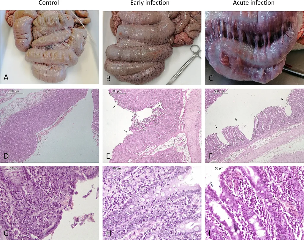 Figure 2. Original Histopathology of Early and Acute Swine Dysentery — Original Figure 2 retained from Pérez-Pérez et al. (2024), CC BY 4.0; doi:10.1186/s40813-024-00375-9. Pig study: uninfected controls n=16, early infection n=8, acute infection n=8; representative images. A/D/G: controls; B/E/H: early; C/F/I: acute. D–I: H&E staining, with original magnifications, scale bars, and arrows preserved. These are pathology images; no new statistical analysis has been added.
Comparison across infection stages shows more pronounced congestion, necrosis, and inflammatory-cell infiltration in the acute group. These original images link colonic lesions to infection stage; they cannot, on their own, establish the incidence of disease across an entire herd.
Why Proliferative Intestinal Mucosa Can Still Reduce Nutrient Utilization
Thickening of the intestinal mucosa in PPE does not imply greater absorptive capacity. Helm and colleagues compared uninfected, infected-unvaccinated, and vaccinated-infected groups of 12 pigs each and found that sucrase-isomaltase transcriptional signals were nearly absent from infected lesions. Together with decreased total-tract nutrient digestibility, the enzyme-localization findings support the interpretation that the lesion contains relatively immature proliferating epithelium rather than a normal functional absorptive surface. [2]
In the same study, average daily gain over days 0–19 after challenge was 1.27, 0.75, and 1.02 kg/day in the respective groups, with significant differences among all three. The orally administered live attenuated vaccine partially improved growth, although performance remained below the uninfected group. Apparent total-tract nitrogen digestibility was 80.3%, 73.9%, and 76.3%, respectively; the difference between the vaccinated-infected and infected-unvaccinated groups was not significant. Growth recovery and individual nutrient-digestion endpoints must therefore be interpreted separately. [2]
Coinfection Creates Interactions Between Two Distinct Intestinal Targets
Daniel and colleagues allocated 45 piglets to coinfection (n=12), Brachyspira only (n=11), Lawsonia only (n=11), and negative control (n=11) groups. Lawsonia was inoculated first, followed by Brachyspira on days 7–9. After day 18, the coinfected group had higher fecal scores than the other groups and more gross large-intestinal lesions. Some tissue endpoints did not differ significantly from the Brachyspira-only group, illustrating endpoint-specific interactions. The authors’ proposed immunosuppression mechanism remains a hypothesis; the principal measured finding was worsened disease under this particular infection sequence. [3]
Microbiota and Diet Jointly Shape Disease Conditions
A 2026 longitudinal field investigation of two farms examined 102 fecal samples from 13 pigs that developed disease and 13 that did not, using metagenomic analysis. Microbial differences were evident before disease onset, although reduced diversity varied by farm. Such longitudinal data are closer to questions of susceptibility than single post-disease samples, but diet, location, and medication history remain important. Relative abundance is not a direct surrogate for microbial function: in Daniel’s coinfection study, the more severely affected coinfected group also had higher relative Lactobacillus abundance than the Brachyspira-only group. [3, 4]
Helm and colleagues compared three diets in 39 individually housed pigs, initially 13 per group, progressively replacing corn distillers dried grains with beet pulp and resistant potato starch. Over 42 days, clinical SD incidence was 85%, 46%, and 15% in the low-, medium-, and high-fermentable-fiber groups (p=0.002). The proportion of culture-positive animals did not differ between groups, showing that disease incidence may fall despite ongoing colonization. The findings support effects of this specific combination of fibers; they do not imply that adding any fiber will yield the same results. [5]
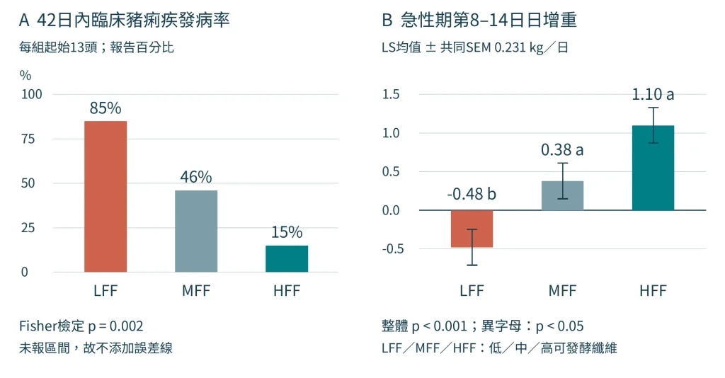 Figure 3. Effects of Specific Fiber Combinations on SD Incidence and Acute-Phase Daily Gain — Redrawn from Helm et al. (2021), Results 3.1 and Table 2; doi:10.3390/ani11020396, CC BY 4.0. Thirty-nine individually housed pigs, initially n=13/group. LFF/MFF/HFF: low/medium/high fermentable fiber diets, progressively substituting beet pulp and resistant potato starch for corn distillers dried grains. A: 0–42-day disease incidence of 85%, 46%, and 15% (Fisher test p=0.002); no confidence intervals reported, so no error bars added. B: days 8–14 ADG of −0.48, 0.38, and 1.10 kg/day; least-squares mean ± pooled SEM 0.231, overall p<0.001; different letters p<0.05. One MFF pig died on day 10 and was included in the original analysis; four LFF pigs received rescue antimicrobial treatment.
Panel A shows how clinical incidence differed by dietary treatment when these ingredients were substituted. Panel B focuses on the acute phase: the low-fermentable-fiber group showed negative daily weight gain, while the medium and high groups performed better. These are two endpoints from one experiment, not independent replications.
What Direct Evidence Exists for Lactobacilli and Cell-Free Supernatants?
Gómez-Martínez and colleagues cocultured 28 anaerobic commensal isolates with B. hyodysenteriae B204. L. amylovorus, Limosilactobacillus mucosae, and Lactiplantibacillus plantarum isolates reduced qPCR-estimated bacterial abundance at 96 hours by approximately 1.86, 1.78, and 1.53 log10/mL, respectively, compared with pathogen-only cultures. These figures describe in vitro growth inhibition, not reduced pathogen shedding on farms. Inhibition kinetics also varied among isolates and time points. [6]
Experiments with cell-free supernatants (CFS) provided further mechanistic clues. Some supernatants, including preparations from the L. mucosae and L. plantarum isolates mentioned above, retained inhibitory activity after neutralization to pH 7.0, indicating that part of the effect was not solely due to culture-acidification. The investigators also quantified lactate and short-chain fatty acids. While these measurements motivate further activity analysis, the principal effector molecule has not been established by single-compound rescue or blocking experiments. For strain development, comparing activity before and after neutralization is more informative than relying on inhibition-zone diameter alone. [6]
Pig Studies of Complex Fermented Preparations Must Preserve Product and Model Identity
The FAM preparation examined by Xu and colleagues contained live lactobacilli, Bacillus spp., and fermentation products; it was a complex probiotic fermented preparation. Twenty-four piglets were assigned to four groups of six: uninoculated control, Lawsonia inoculation, FAM plus inoculation, and vaccine plus inoculation. The authors identified the inoculation material as the commercial attenuated Enterisol Ileitis vaccine and sampled on day 11 after inoculation. These outcomes belong to that specific inoculation model and should not be conflated with challenges using virulent field isolates. [7]
Compared with inoculated untreated pigs, the FAM group showed better daily weight gain, lower ileal Lawsonia RNA signal, more goblet cells and MUC2 expression, and improvements in serum markers associated with intestinal permeability. Biologically, this indicates that pathogen localization, mucus barrier responses, and growth can change together, but any effect must be attributed to the whole FAM preparation. Only one pen per treatment group was used, so pen and dietary-treatment effects could not be separated, limiting causal inference for production performance. [7]
Table 1. Materials and Measured Endpoints in Key Studies
| Material and source | Study setting | Main findings and interpretation |
|---|---|---|
| Commensal lactobacilli and CFS [6] | In vitro coculture; supernatant concentration and pH comparisons; three replicates per condition | Lower qPCR-estimated pathogen abundance; activity partly retained after neutralization; no farm-level efficacy study |
| FAM complex live-bacteria fermented preparation [7] | 24 pigs; four groups of six, one pen per group; inoculation with attenuated vaccine material | Growth, RNA signal, goblet cells, and MUC2 improved; component and pen effects cannot be isolated |
| P/S organic acids; D botanical; F fungal-fermented rye [8] | Ex vivo colon; 10 donor pigs for Brachyspira, 5 for Lawsonia | Epithelial/inflammatory responses varied by material, time, and pathogen; these are not one postbiotic category |
| Bacillus-derived surfactin [9] | In vitro inhibition and electron microscopy; three experimental replicates | Morphological damage and growth inhibition; no same-study demonstration of reduced shedding in pigs |
Compiled from original methods and results. Individual pigs, pens, donor tissues, and culture replicates are different experimental units.
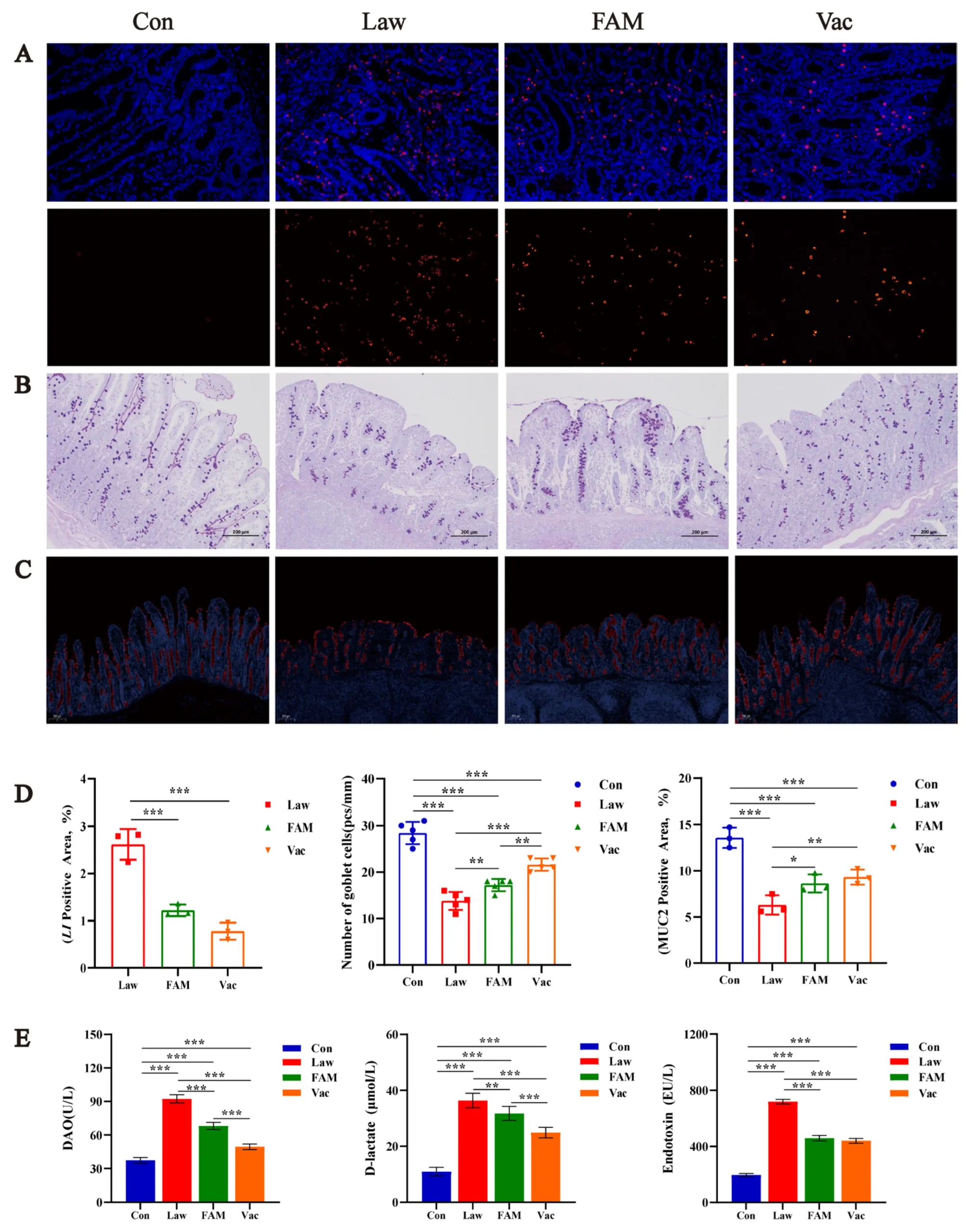 Figure 4. Lawsonia RNA Signal and Mucosal Markers in the FAM Model — Original Figure 6 from Xu et al. (2023); doi:10.3390/biology12060879, CC BY 4.0. Twenty-four piglets, n=6 per group, one pen per group: Con, untreated and uninoculated; Law, inoculated; FAM, fermented preparation plus inoculation; Vac, vaccine plus inoculation. The inoculum was a commercial attenuated vaccine. A: Lawsonia RNA in situ hybridization (red positive signal, blue DAPI); B: PAS-stained goblet cells; C: MUC2; D: signal area and goblet-cell quantification; E: DAO, D-lactate, and endotoxin. Scale bars in B/C are 200 µm. The authors report mean ± SD; *p<0.05, **p<0.01, ***p<0.001. Three or six points appear in some D/E measures; the relationship between subsamples and microscopic fields is incompletely documented and should not be uniformly called n=6 for each plot.
In panels A/D, the FAM group showed less Lawsonia RNA signal than the Law group; in panels B–D, goblet-cell and MUC2 outcomes were improved, although not all reached control levels. Panel E shows serum permeability-associated markers rather than direct measurement of whole-intestine permeability. Together, these images and measurements support mucosa-associated effects of the complex preparation in this specific inoculation model.
Epithelial Protection and Direct Antibacterial Activity Are Distinct Mechanisms
In an ex vivo pig-colon study, de Groot and colleagues distinguished pathogen-only exposure, material-only exposure, and simultaneous exposure. For Brachyspira, the P preparation containing short- and medium-chain fatty acids reduced mucus thickness under some conditions and was associated with better epithelial coverage or lower TNF-alpha signal. Carob and thyme extracts and fungal-fermented rye showed different responses. In particular, the fungal-fermented material did not act consistently across Lawsonia and Brachyspira contexts, emphasizing the importance of pathogen, comparator, and sampling time. [8]
Donor animals and tissue pieces must be counted separately: Lawsonia experiments used five donors, while the Brachyspira experiments involved ten donors because materials were evaluated in batches. Multiple tissue pieces from one pig are not equivalent to multiple independent pigs. The original figure also used a double asterisk for p=0.06, which should be interpreted as a trend under the stated alpha=0.05 threshold. Separately, Horng and colleagues observed Brachyspira cell-shape damage in antibacterial assays and scanning electron microscopy with surfactin. This is in vitro evidence of direct effects on bacterial cells; surfactin is a Bacillus-derived lipopeptide, not a lactobacillus preparation. [8, 9]
Postbiotic Terminology and the Role of Integrated Disease Control
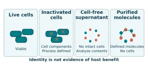
According to the ISAPP consensus, a postbiotic is a preparation of inanimate microorganisms and/or their components that confers a health benefit on the host, potentially with accompanying metabolites. Live-bacteria-containing FAM, cell-free supernatants, and purified fatty acids should each be identified by their actual material type; botanical extracts, mineral ingredients, and host-defense peptides belong to separate categories. Precise terminology preserves information about the source of activity and manufacturing process and improves comparisons among strains, fermentation products, and postbiotics. [10]
Table 2. Terminology and Functional Levels of Research Materials
| Research material | Information that must be preserved | Main research question |
|---|---|---|
| Live bacteria and complex fermented preparations | Strain, viable count, fermentation substrate, coexisting components | Effects of the whole preparation in the stated model |
| Cell-free supernatants | Bacteria-removal method, concentration, pH, medium controls | Relationship between soluble inhibitory activity and acidification |
| Preparations meeting the postbiotic definition | Inactivation method, microbial cells/components, demonstrated host benefit | Health effect of a specific inanimate microbial preparation |
| Purified metabolites and other materials | Chemical identity, origin, purity; separate botanical/mineral/host sources | Direct function of a defined molecule or material |
Classification is based on reference [10] and the material identities discussed here. A supernatant or metabolite does not automatically qualify as a postbiotic because it originates from microorganisms. [10]
Field evidence provides a separate benchmark for clinical endpoints. In a randomized, blinded trial involving 3,261 pigs, Lawsonia-associated mortality was 0.45% in the intradermal inactivated-vaccine group versus 1.65% in controls (p=0.0022). This mortality outcome relates to that specific vaccine and field setting; it addresses a different question from the digestibility, growth, or fermented-preparation experiments above and should be interpreted within its own comparison and endpoints. [11]
Taken together, the most relevant research directions for lactobacilli and fermentation-derived materials include competition with pathogens, soluble inhibitory activity, mucus and epithelial function, and dietary modulation of disease expression. Practical disease control still requires pathogen diagnosis, nutrition, vaccination, and biosecurity as an integrated framework. Material-specific findings are the basis for evidence that can be meaningfully compared and reproduced.
Frequently Asked Questions
Is every fermented preparation a postbiotic? Classification depends on viability, cellular components, and preparation details. FAM study results concern a complex fermented preparation that contains live bacteria. [7, 10]
Does lower disease incidence mean that Brachyspira has been eliminated? In Helm’s experiment, incidence fell but the proportion of culture-positive animals did not differ among groups. These outcomes require separate interpretation. [5]
Image Sources and Usage Notes
Figure 1 is a BioRender conceptual illustration; Figure 2 reproduces the original Figure 2 from reference [1]; Figure 3 was reconstructed from the findings and Table 2 in reference [5]; Figure 4 reproduces original Figure 6 from reference [7]. Figures 2–4 are attributed under CC BY 4.0. See the captions and references for the original figures, data tables, and DOIs.
References & Notes
- [1] Pérez-Pérez, Lucía; Carvajal, Ana; Puente, Héctor; Peres Rubio, Camila et al. (2024). New insights into swine dysentery: faecal shedding, macro and microscopic lesions and biomarkers in early and acute stages of Brachyspira hyodysenteriae infection. Porcine Health Management 10(1):24. doi:10.1186/s40813-024-00375-9
- [2] Helm, Emma T.; Burrough, Eric R.; Leite, Fernando L.; Gabler, Nicholas K. (2021). Lawsonia intracellularis infected enterocytes lack sucrase-isomaltase which contributes to reduced pig digestive capacity. Veterinary Research 52(1):90. doi:10.1186/s13567-021-00958-2
- [3] Daniel, Amanda G. S.; Pereira, Carlos E. R.; Dorella, Fernanda; Pereira, Felipe L. et al. (2023). Synergic Effect of Brachyspira hyodysenteriae and Lawsonia intracellularis Coinfection: Anatomopathological and Microbiome Evaluation. Animals 13(16):2611. doi:10.3390/ani13162611
- [4] Pérez-Pérez, Lucía; Arguello, Héctor; Cobo-Díaz, José F.; Galisteo, Cristina et al. (2026). From predisposition to recovery: field evidence of interactions between the gut microbiota and Brachyspira hyodysenteriae infection. Veterinary Research 57(1):25. doi:10.1186/s13567-025-01646-1
- [5] Helm, Emma T.; Gabler, Nicholas K.; Burrough, Eric. R. (2021). Highly Fermentable Fiber Alters Fecal Microbiota and Mitigates Swine Dysentery Induced by Brachyspira hyodysenteriae. Animals 11(2):396. doi:10.3390/ani11020396
- [6] Gómez-Martínez, Samuel; Pérez-Pérez, Lucía; Ucero-Carrretón, Alejandro; López-García, Álvaro et al. (2026). Exploring the potential for competitive exclusion of commensal probiotic candidates against the insidious swine pathogen Brachyspira hyodysenteriae. Animal Microbiome 8(1):19. doi:10.1186/s42523-025-00511-y
- [7] Xu, Tingting; Guo, Yong; Zhang, Yuanyuan; Cao, Kai et al. (2023). Alleviative Effect of Probiotic Ferment on Lawsonia intracellularis Infection in Piglets. Biology 12(6):879. doi:10.3390/biology12060879
- [8] de Groot, Nienke; Meneguzzi, Mariana; de Souza, Barbara; de O. Costa, Matheus (2022). In Vitro Screening of Non-Antibiotic Components to Mitigate Intestinal Lesions Caused by Brachyspira hyodysenteriae, Lawsonia intracellularis and Salmonella enterica Serovar Typhimurium. Animals 12(18):2356. doi:10.3390/ani12182356
- [9] Horng, Yi-Bing; Yu, Yu-Hsiang; Dybus, Andrzej; Hsiao, Felix Shih-Hsiang et al. (2019). Antibacterial activity of Bacillus species-derived surfactin on Brachyspira hyodysenteriae and Clostridium perfringens. AMB Express 9(1):188. doi:10.1186/s13568-019-0914-2
- [10] Salminen, Seppo; Collado, Maria Carmen; Endo, Akihito; Hill, Colin et al. (2021). The International Scientific Association of Probiotics and Prebiotics (ISAPP) consensus statement on the definition and scope of postbiotics. Nature Reviews Gastroenterology & Hepatology 18(9):649–667. doi:10.1038/s41575-021-00440-6
- [11] Jacobs, A. A. C.; Harks, F.; Pauwels, R.; Cao, Q. et al. (2020). Efficacy of a novel intradermal Lawsonia intracellularis vaccine in pigs against experimental infection and under field conditions. Porcine Health Management 6(1):25. doi:10.1186/s40813-020-00164-0
- [12] Lin, Susanne Je-Han ; Helm, Emma T. ; Gabler, Nicholas K. ; Burrough, Eric R. (2023). Acute infection with Brachyspira hyodysenteriae affects mucin expression, glycosylation, and fecal MUC5AC. Frontiers in Cellular and Infection Microbiology 12:1042815. doi:10.3389/fcimb.2022.1042815
- [13] Hakimi, Maria; Ye, Fangshu; Stinman, Chloe C.; Sahin, Orhan et al. (2024). Antimicrobial susceptibility of U.S. porcine Brachyspira isolates and genetic diversity of B. hyodysenteriae by multilocus sequence typing. Journal of Veterinary Diagnostic Investigation 36(1):62–69. doi:10.1177/10406387231212189
This is a narrative scientific review for research and technical exchange. Diagnosis and control in individual cases must be based on the actual pathogens and farm conditions.