
After coccidial infection, oocyst shedding, intestinal lesions and barrier recovery describe different aspects of parasite output, tissue injury and host function. These outcomes can be related without changing in parallel. Chicken studies show that the microbial background can influence lesion severity, while lactobacilli and other microbial materials may affect tight junctions and inflammatory responses. Interpreting this evidence requires a framework that connects the parasite, the microbiota and the epithelium while retaining the distinct meaning of each endpoint.[1, 2, 3, 4]
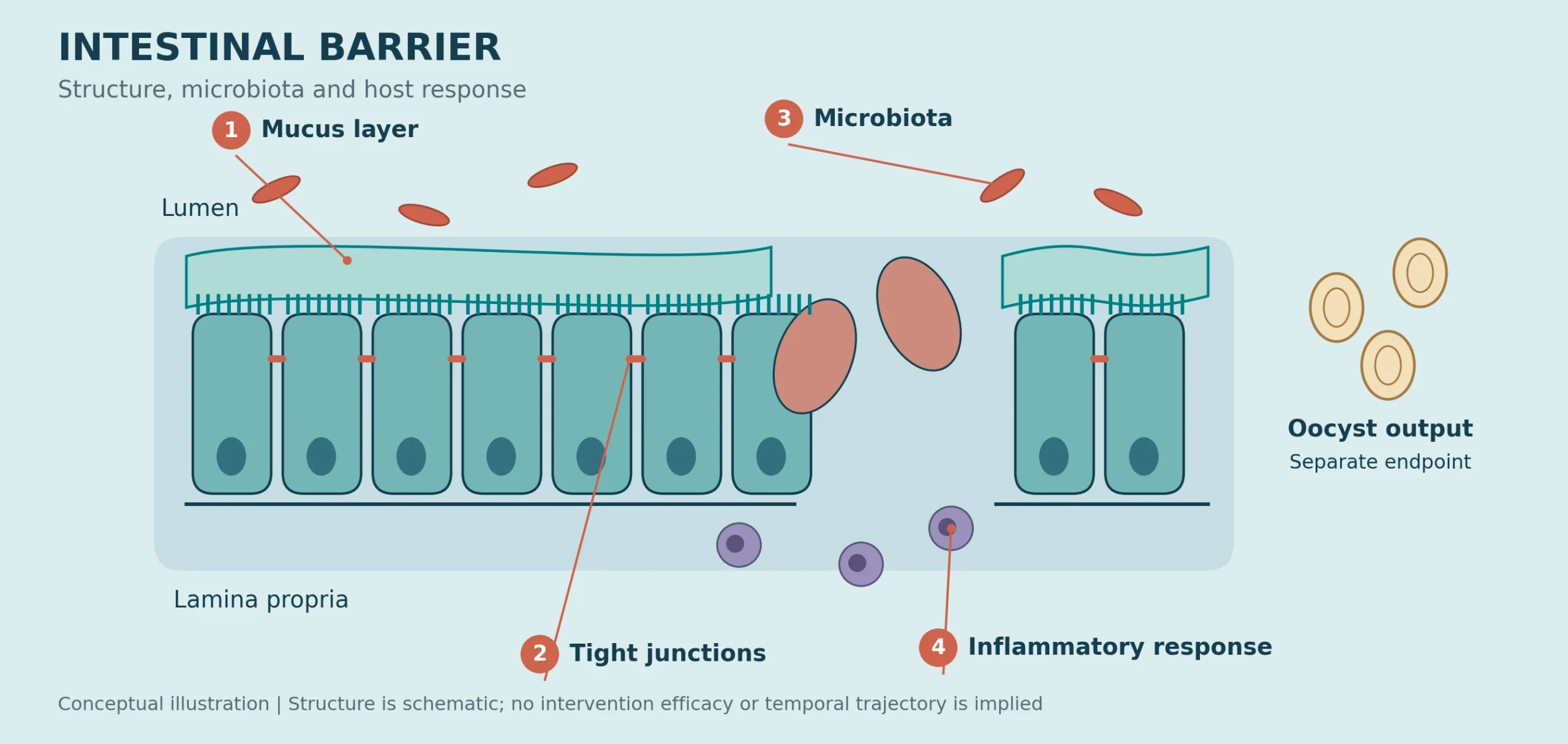
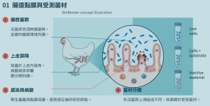
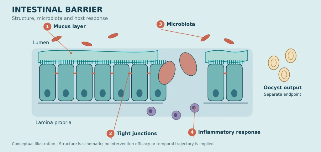
Conceptual illustration of the intestinal mucosa and microbiota, created as an original HYGEM vector graphic. This is the conceptual portion of the EDM, not a histological image or an empirical efficacy figure.[1, 2, 3]
From mucus to epithelium: coccidia disrupt an integrated barrier
Different Eimeria species preferentially affect different intestinal regions in poultry. E. acervulina primarily involves the duodenum and proximal small intestine, E. maxima the mid-small intestine, and E. tenella the ceca. Infection damages epithelial cells, shortens or disrupts villi, and is accompanied by local inflammation. The same fecal oocyst reading can therefore occur against different anatomical and tissue-injury backgrounds; it cannot be translated directly into the same level of absorptive function.[1]
The mucus layer, goblet cells and tight junctions together maintain the interface between the intestinal lumen and underlying tissue. In their E. tenella study, Choi and colleagues found lower cecal goblet cell numbers and lower mucus concentrations in the intestinal contents, while MUC2 transcription showed a nonlinear response. Mucin gene signals may reflect compensation rather than the formation of continuous, intact mucus coverage. Interpretation of this chemical barrier therefore also requires information on goblet cells and the actual mucus condition.[2]
Table 1. Four measurement levels in intestinal barrier research
| Measurement level | Main data | Question addressed | Interpretation |
|---|---|---|---|
| Parasite output | Fecal OPG / total oocyst output | Has output changed? | Different times and denominators are not interchangeable |
| Mucus and tissue | Goblet cells, mucus and morphology | Are coverage and epithelium damaged? | MUC2 mRNA is not a secretion measurement [2] |
| Junction components | ZO-1, CLDN and Occludin | How do transcripts and proteins change? | Expression direction alone does not establish function [1, 3] |
| Function | FITC-d and indirect serum markers | What is the leakage / barrier status? | Separate direct permeability from indirect markers [1, 4] |
The microbiota also contributes to lesion development
Tomal and colleagues compared conventional chickens, germ-free chickens and germ-free chickens in which microbiota had been reintroduced. At seven days after infection, under conditions with similar parasite loads, germ-free chickens had fewer cecal lesions. Reintroducing microbiota from conventional chickens increased lesions and mucosal damage. By changing the microbial background, this study directly supports microbial involvement in infection pathology rather than inferring an association from sequencing data alone.[3]
The response in conventional chickens included inflammatory cell infiltration and higher inflammation-related signals such as IL18 and PTGS2. Enterobacteria were also detected more frequently in the spleen: in 4/7 infected conventional chickens, compared with 1/11 uninfected conventional chickens. This small-sample observation of bacterial translocation is not a farm-level secondary-infection rate. It connects epithelial disruption with the risk of luminal bacteria crossing the barrier. Germ-free models help identify mechanisms; these findings do not support a farming strategy that removes the entire intestinal microbiota.[3]
Permeability can increase without a significant decline in junction-gene expression
In the mixed-Eimeria infection study by Graham and colleagues, mean serum fluorescein isothiocyanate–dextran (FITC-d) increased from 34.22 ng/mL in uninfected chickens to 229.50 ng/mL in infected chickens at six days after infection, indicating increased intestinal permeability. However, mean relative ileal ZO-1 and OCLN mRNA levels were each 0.88 times the control value, with no statistically significant difference between groups. Function had changed without a corresponding significant change in these individual transcriptional indicators, demonstrating the need for assessment at several biological levels.[1]
Tight-junction function also depends on protein abundance, intracellular localization, junctional continuity and epithelial integrity. CLDN1 in the Tomal study provides another example: severe lesions were accompanied by increased transcription rather than a uniform decrease. Increased expression of a tight-junction gene cannot invariably be interpreted as protection, and a nonsignificant difference cannot establish that barrier function is normal.[1, 3]
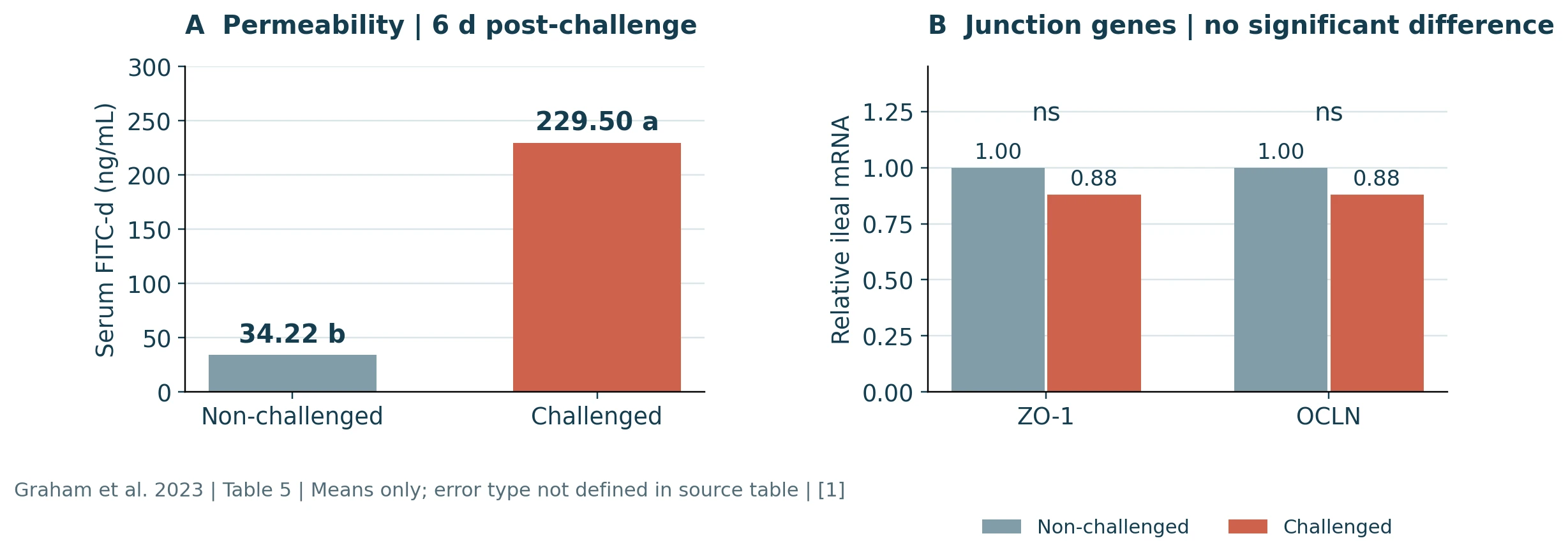
Independently plotted from the means in Graham et al. (2023), Table 5. Left: serum FITC-d; right: relative ileal mRNA. Sampling was at six days after infection. The study initially had five uninfected pens and seven infected pens. The FITC-d methods specify four birds per pen; gene-expression sampling was n = 8 per group. The source table does not clearly define the type of error represented by ±, so only means are plotted and no error bars have been added. Different letters indicate p < 0.05 between groups; ns denotes a nonsignificant difference. CC BY 4.0.[1]
Serum FITC-d increased, whereas ZO-1 and OCLN did not differ significantly between groups. An individual mRNA indicator cannot replace a functional measurement.
P8: shedding, lesions and barrier indicators show different responses
The Lactobacillus plantarum P8 study by Wang and colleagues included an uninfected control, an infected control, two P8 groups and a diclazuril group. At five days after infection, oocysts per gram of excreta (OPG) were 11.96 × 10⁵/g in the infected control, compared with 4.52, 1.28 and 0.55 × 10⁵/g in P8L, P8H and the drug group, respectively. P8H and the drug group shared a statistical letter for OPG. Their cecal lesion scores, however, were 1.27 and 0.40, respectively, and differed significantly; the scores were 1.09 for P8L and 2.36 for the infected control. Similar statistical groupings for parasite output did not produce the same tissue-lesion outcome.[4]
At 21 days of age, mean relative jejunal Occludin protein expression increased from 0.380 in the infected control to 0.761 in P8H, while serum D-lactate decreased from 77.82 to 56.24 μmol/L. At 42 days of age, Occludin values were 0.548 in P8H and 0.282 in the infected control. Although P8H still shared a statistical letter with the uninfected group, this does not demonstrate equivalence. The protein and indirect leakage indicators support an improvement in host barrier status, without directly demonstrating that every aspect of permeability had recovered.[4]
P8 did not reverse all immune and microbiota indicators. At 21 days of age, IL-10 did not return to the uninfected level with P8, and several microbiota-diversity indicators did not improve significantly. Different host responses therefore have different magnitudes of change. The percentage reduction in oocyst output cannot stand in for the extent of overall intestinal recovery.[4]
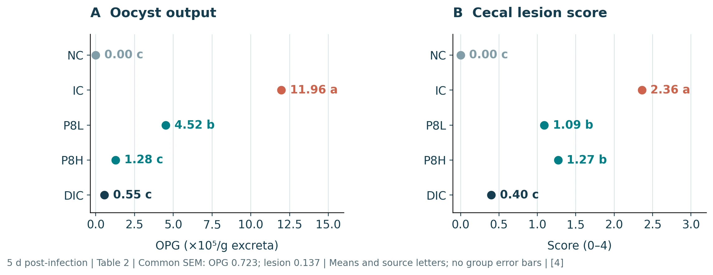
Independently plotted from Wang et al. (2021), Table 2, at five days after infection. NC = uninfected control; IC = infected control; P8L and P8H = the two P8 groups in the original study; DIC = diclazuril. Each group initially had 10 cages with 10 birds per cage. Excreta were collected by cage; cecal lesion assessment used n = 6 samples per group. Common SEM values in the source table were 0.723 for OPG (×10⁵/g) and 0.137 for lesions. These common statistics have not been converted into individual group error bars. Letters reproduce the original multiple-comparison groupings: means without a shared letter differ at p < 0.05. The overall p-value was 0.001 for each endpoint. CC BY 4.0.[4]
P8H and DIC share letter c for OPG but have letters b and c, respectively, for cecal lesions. A shared letter indicates no statistically significant difference; it does not demonstrate equivalence.
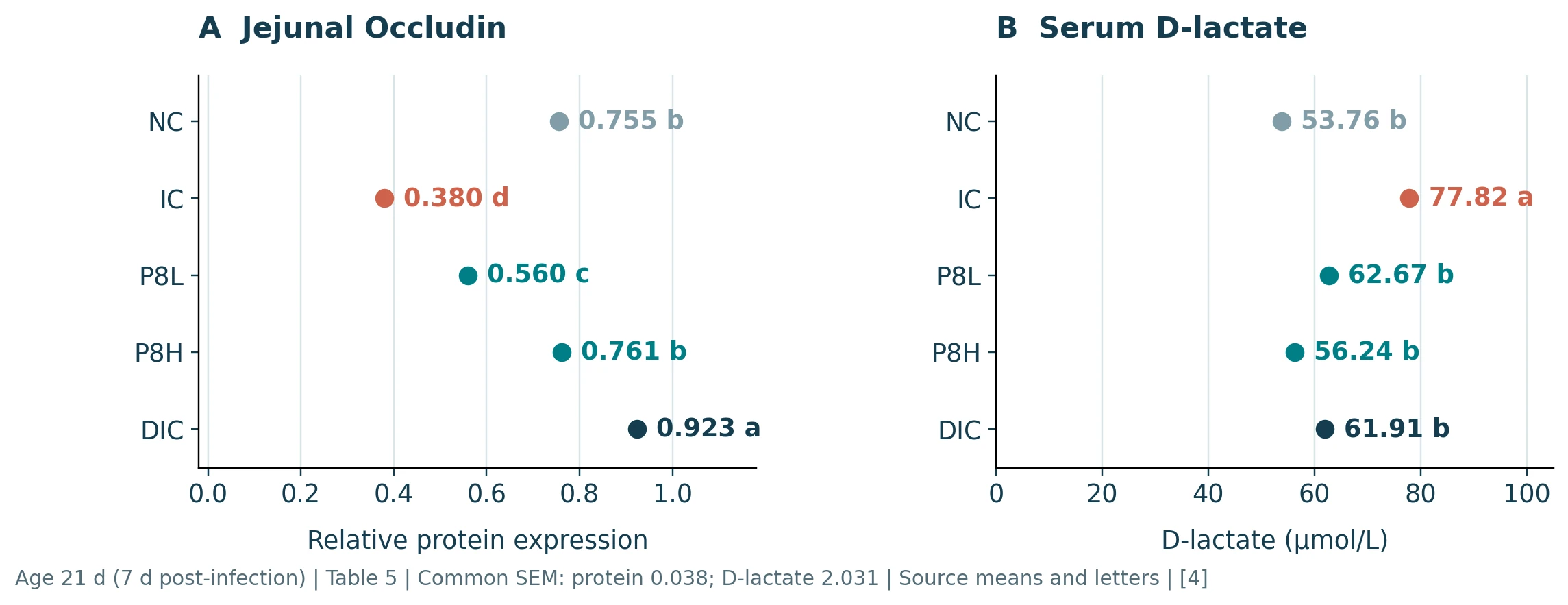
Independently plotted from Wang et al. (2021), Table 5, at 21 days of age, or seven days after infection. Groups are defined in Figure 2. Blood and intestinal sampling used one bird from each of 10 cages per group; Western blot analysis used jejunal mucosa from n = 6 per group. Protein values are relative expression as defined in the original study; serum D-lactate is expressed in μmol/L. Common SEM values were 0.038 for Occludin and 2.031 for D-lactate; individual group error bars have not been added. Means without a shared letter differ at p < 0.05. The overall p-values were reported as 0.000 for Occludin, presented here as p < 0.001, and 0.001 for D-lactate. CC BY 4.0.[4]
Occludin expression and D-lactate in P8H shifted toward the uninfected values. D-lactate is an indirect marker and is not interchangeable with FITC-d permeability.
BS22 adds evidence at the protein and tissue levels
The 2026 BS22 study examined co-infection with E. tenella and E. acervulina. Each group initially contained 18 chickens, with duodenal and cecal comparisons at five and ten days after infection. The strain-treated group had less tissue damage and better preservation of ZO-1 and CLDN5 mRNA and ELISA-measured protein levels than the infected group. Consistent changes across several biological levels strengthen the interpretation of barrier protection.[5]
The functional scope of this evidence is nevertheless defined by what was measured: tissue morphology and protein content are not direct tracer-permeability measurements. The original paper also describes the units of its oocyst data inconsistently. This review therefore does not convert those values or rank them against P8 OPG data. Together, the P8 and BS22 findings show that specific strains can influence host responses; they do not establish the same effect for all L. plantarum strains.[4, 5]
Synbiotics and postbiotic materials: lesion improvement does not establish full functional recovery
The synbiotic studied by Shah and colleagues contained Limosilactobacillus reuteri, Enterococcus faecium, Bifidobacterium animalis, Pediococcus acidilactici and fructooligosaccharides. In a necrotic-enteritis model involving E. maxima and Clostridium perfringens, the synbiotic reduced mid-intestinal lesions at 21 days of age but did not improve FITC-d permeability at that time; the synbiotic main-effect p-value was 0.98. This is an example of pathology and function responding differently. Because the model involved a combined infection, the findings cannot be attributed entirely to activity against coccidia alone.[6]
In the coccidia study by Alagbe and colleagues, enzymatically treated yeast was associated with lower oocyst counts, higher OCLN expression and greater ileal goblet cell density, but did not fully offset the adverse effects of infection.[7] A separate comparison of viable lactobacilli, heat-inactivated lactobacilli and culture supernatant used a complex model involving material from a coccidiosis vaccine, an infectious-bursal-disease vaccine background, C. perfringens and deoxynivalenol (DON). The heat-inactivated material showed some tissue protection, but this cannot be treated as direct efficacy evidence for uncomplicated coccidiosis.[8]
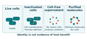
The postbiotic consensus requires a preparation of inanimate microorganisms and/or their components that confers a health benefit. Purified metabolites or sterile supernatants do not automatically fall into the same category merely because they originate from fermentation.[9] Barrier studies should retain the identity of the material actually tested before determining whether the evidence concerns mucus, inflammation, tissue structure or function.
Connecting parasite control with measured barrier recovery
Lower oocyst output can coexist with less severe lesions, but the two endpoints are not interchangeable. Epithelial recovery also requires mutually supporting evidence from goblet cells, junction proteins and permeability measurements. The available studies show that the consequences of coccidial infection reflect the combined contributions of the parasite, the microbial background and the host response. Some microbial materials can improve selected components of this interaction.[1, 2, 3, 4, 5, 6, 7, 8]
When assessing evidence for application, first align the intestinal region, sampling time and control group, then connect shedding, morphology, protein and functional outcomes. Studies based on cross-sectional sampling alone cannot establish faster recovery. This approach retains the research value of individual strains and preparations while keeping the term “gut recovery” tied to measured outcomes.
Table 2. Microbial materials and study contexts
| Study context | Main barrier evidence | Interpretation |
|---|---|---|
| P8 / mixed Eimeria [4] | Jejunal proteins, D-lactate and pathology | Keep host and parasite endpoints separate |
| BS22 / two Eimeria species [5] | ZO-1 and CLDN5 mRNA / proteins and tissue | Supports tissue protection; permeability not measured directly |
| Synbiotic / necrotic enteritis [6] | Mid-intestinal lesions, FITC-d and immune responses | Lesion improvement and functional recovery may diverge |
| Yeast or inactivated materials [7, 8] | Goblet cells, OCLN or tissue protection | Interpret within the material and infection context tested |
Further Reading
-
Microbiome Nutrition and Intestinal Barrier Function in Livestock
-
How Lactobacillus plantarum Helps Restore the Intestinal Barrier in Chicks Challenged by Salmonella
References
[1] Graham Danielle, Petrone-Garcia Victor M, Hernandez-Velasco Xochitl et al. (2023). Assessing the effects of a mixed Eimeria spp. challenge on performance, intestinal integrity, and the gut microbiome of broiler chickens. Frontiers in Veterinary Science 10: 1224647. doi:10.3389/fvets.2023.1224647
[2] Choi Janghan, Kim Wookyun, Acevedo-Villanueva Keila (2022). Interactions of Microbiota and Mucosal Immunity in the Ceca of Broiler Chickens Infected with Eimeria tenella. Vaccines 10(11): 1941. doi:10.3390/vaccines10111941
[3] Tomal Florian, Sadrin Guillaume, Gaboriaud Pauline et al. (2023). The caecal microbiota promotes the acute inflammatory response and the loss of the intestinal barrier integrity during severe Eimeria tenella infection. Frontiers in Cellular and Infection Microbiology 13: 1250080. doi:10.3389/fcimb.2023.1250080
[4] Wang Yang, Lv Xiaoguo, Li Xuemin et al. (2021). Protective Effect of Lactobacillus plantarum P8 on Growth Performance, Intestinal Health, and Microbiota in Eimeria-Infected Broilers. Frontiers in Microbiology 12: 705758. doi:10.3389/fmicb.2021.705758
[5] Zhang Xinhui, Zhou Xuan, Wang Lidan et al. (2026). Protective efficacy of Lactobacillus plantarum BS22 against Eimeria tenella and Eimeria acervulina co-infection in broiler chickens. Poultry Science 105(10): 107342. doi:10.1016/j.psj.2026.107342
[6] Shah Bikas R, Hakeem Walid A, Shanmugasundaram Revathi et al. (2023). Effect of synbiotic supplementation on production performance and severity of necrotic enteritis in broilers during an experimental necrotic enteritis challenge. Poultry Science 102(10): 102959. doi:10.1016/j.psj.2023.102959
[7] Alagbe Emmanuel Oluwabukunmi, Schulze Hagen, Adeola Olayiwola (2023). Growth performance, nutrient digestibility, intestinal morphology, cecal mucosal cytokines and serum antioxidant responses of broiler chickens to dietary enzymatically treated yeast and coccidia challenge. Journal of Animal Science and Biotechnology 14: 57. doi:10.1186/s40104-023-00846-z
[8] de Souza Marielen, Baptista Ana Angelita Sampaio, Menck-Costa Maísa Fabiana et al. (2024). Modulation of Broiler Intestinal Changes Induced by Clostridium perfringens and Deoxynivalenol through Probiotic, Paraprobiotic, and Postbiotic Supplementation. Toxins 16(1): 46. doi:10.3390/toxins16010046
[9] Salminen S, Collado MC, Endo A et al. (2021). The International Scientific Association of Probiotics and Prebiotics (ISAPP) consensus statement on the definition and scope of postbiotics. Nature Reviews Gastroenterology & Hepatology 18: 649–667. doi:10.1038/s41575-021-00440-6
Figure sources: the three data figures were independently plotted from the source-table means and statistical information. The conceptual graphic is an original schematic. Literature search cutoff for the Chinese source article: October 22, 2026.