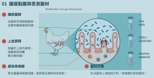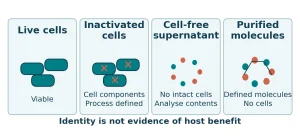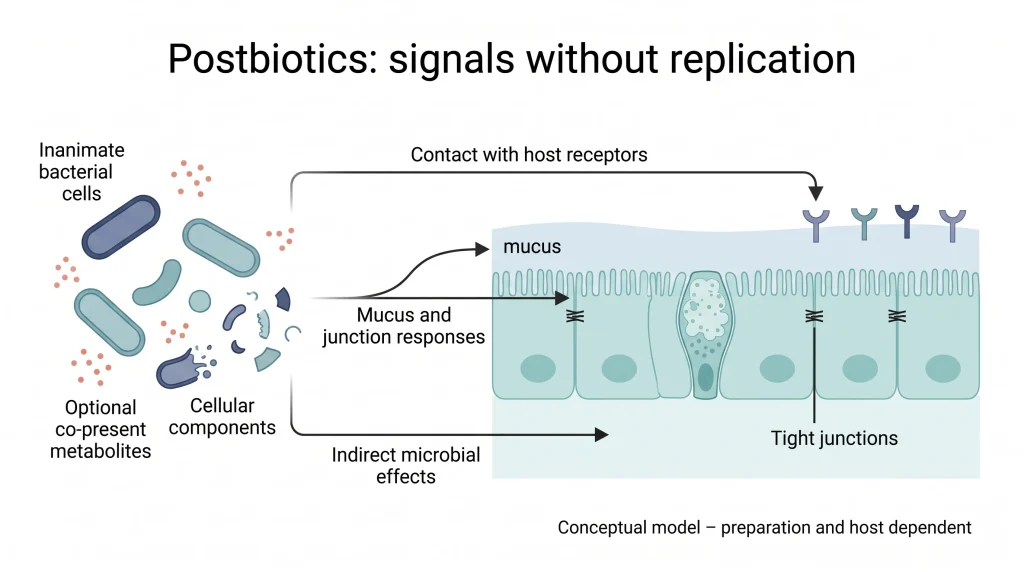

Postbiotics are scientifically relevant because some microbial structures and associated biological activities can interact with a host after the microorganisms have lost their ability to replicate. Colonization is not required for cell envelopes, surface structures or other constituents in a preparation to influence mucus, epithelial junctions or immune responses. However, the absence of live cells describes a material state; a health benefit must be established for the tested preparation in a defined host. Understanding postbiotics therefore requires a connected account of material identity, processing and measured outcomes. [1, 2]
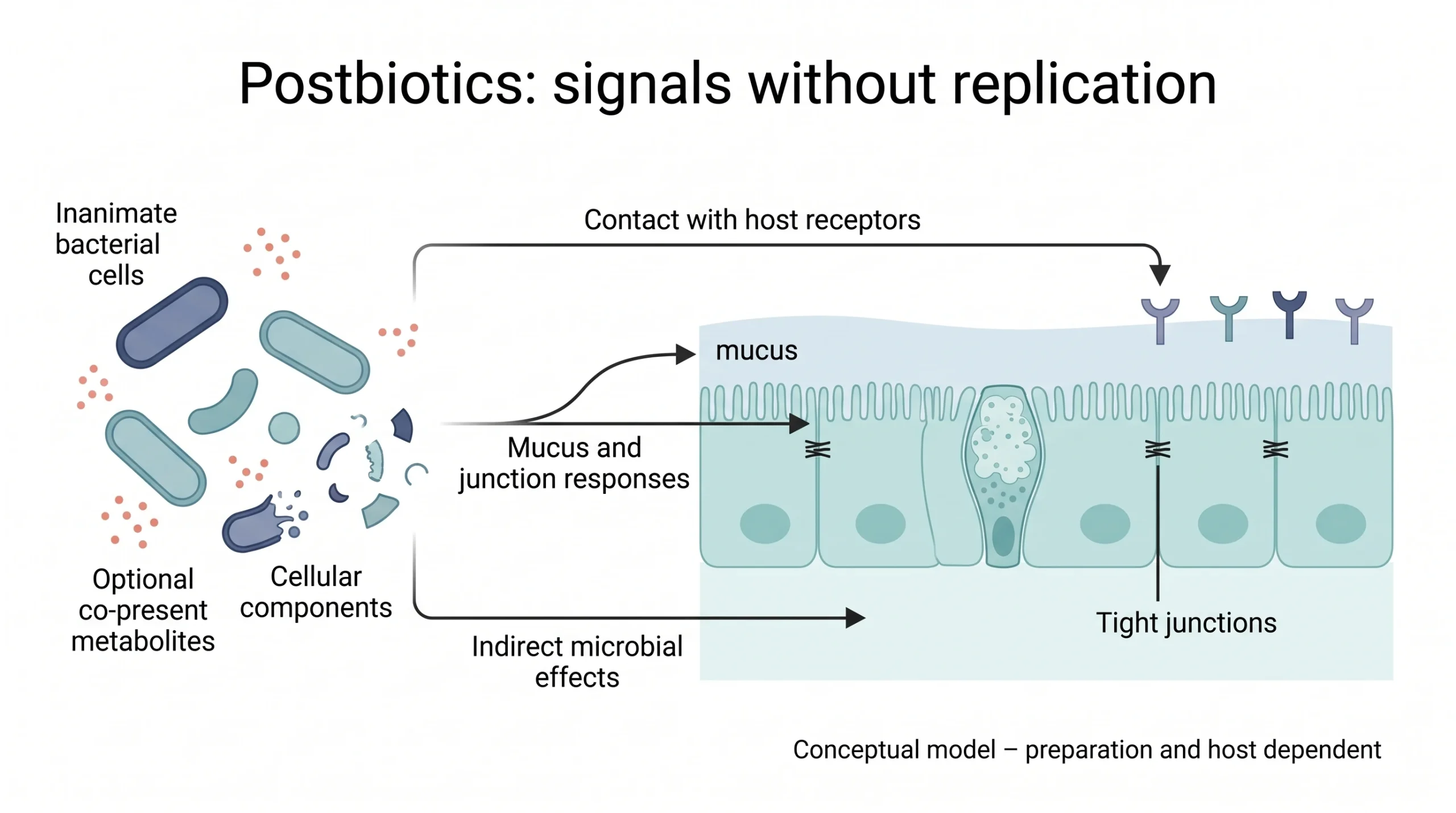
Inanimate cells, cellular components and optional co-present metabolites may participate in intestinal responses through direct host contact or indirect routes. This is a conceptual illustration without experimental groups, sample sizes, axes or statistics; it does not assign every depicted effect to every preparation. Created with BioRender, informed by the ISAPP consensus. [1, 2]
Read from the material on the left toward the host interface. Arrows represent a general framework, rather than a single established causal chain assembled from different preparations.
What a postbiotic preparation contains
The 2021 ISAPP consensus defines a postbiotic as a preparation of inanimate microorganisms and/or their components that confers a health benefit on the host. The defining elements are a microbial origin, inanimate cells or cellular components, and a demonstrated health benefit. The source microorganism does not first have to qualify as a probiotic. Purified lactate, butyrate or vitamins, and filtrates without relevant cellular constituents, do not automatically qualify simply because they originate from fermentation. [1, 2]
This distinction is useful when reading earlier literature. Some studies used the term postbiotics for secreted proteins, culture supernatants and purified metabolites. These studies can still explain microbial effector molecules, but the identity of the tested material must remain explicit. Inactivated biomass, fermentation preparations containing cells, cell-free supernatants and purified proteins have different compositions; a shared label does not make their effects interchangeable. [1, 2, 3]
Table 1. Identifying microbial materials
| Material | Key characteristics | Classification in this article |
|---|---|---|
| Inanimate cells or cellular components | Defined preparation and composition, with a host health benefit | Can meet the adopted postbiotic definition [1, 2] |
| Cell-free supernatant | Check relevant cellular constituents and health-benefit evidence | Absence of live cells is not sufficient [1, 2] |
| Purified organic acid or secreted protein | Isolated molecule or metabolite | Use its material name; not automatically a postbiotic [1, 2, 3] |
| Inactivated fermentation mixture | Biomass, matrix and other constituents remain together | Attribute the effect to the complete tested preparation [2, 5] |
Why biological activity can remain after inactivation
Hosts recognize molecular structures, not only whether a microorganism can reproduce. Inanimate cells may retain surface features that interact with host receptors, while a preparation may also contain cell fragments and co-present metabolites. Effects may involve direct contact with the host or indirect changes mediated by the intestinal environment and resident microbiota. Neither route requires the inactivated organism to resume growth. [1, 2]
Cellular structures and soluble molecules provide different mechanistic clues. Gao and colleagues observed increased mucus production when Caco-2 cells were exposed to a Lactobacillus rhamnosus GG culture supernatant. Protease treatment weakened this response, whereas nuclease and carbohydrate-digesting enzyme treatments did not show the same pattern. The researchers then identified and produced the HM0539 protein and examined mucin and barrier function, moving from an active fermentation preparation toward a more specific source of activity. [3]
The HM0539 study predates the 2021 consensus. Here, its purified protein is classified as a microbial effector molecule rather than equated with a complete postbiotic preparation. The enzyme comparisons in the original Figure 1 support a protein contribution to the mucus response induced by the supernatant. They do not establish that all lactobacillus supernatants contain the same protein or that every inactivation process preserves the effect. [1, 2, 3]
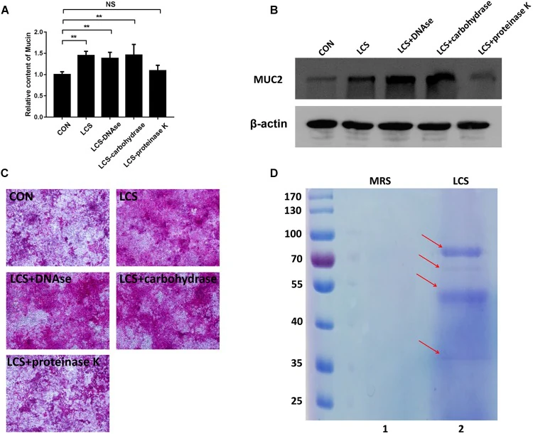
Gao et al. (2019), original Figure 1, reproduced in full. Material: LGG culture supernatant (LCS); model: Caco-2 cells. CON is the control; other groups received LCS or LCS treated with DNase, carbohydrate-digesting enzymes or proteinase K. A: relative mucin content; B: MUC2 immunoblot with β-actin loading control; C: PAS staining; D: SDS-PAGE of supernatant proteins. Means ± SEM; **p < 0.01 and NS, not significant, apply only to bracketed comparisons. The original legend does not specify n or the independent experimental unit for this figure; repetition counts from other assays are not substituted. No scale bar is supplied for C. CC BY. [3]
Protease treatment weakens the mucin response, unlike the other enzyme treatments. This supports a protein contribution to supernatant activity and provided a lead for identifying HM0539. The figure is not an efficacy test of intact inactivated cells.
Barrier protection requires functional measurements
The intestinal barrier includes mucus, epithelial cells and intercellular junctions. Increased MUC2 or tight-junction protein expression provides information about structure and regulation. Transepithelial electrical resistance (TEER) and tracer permeability add functional evidence of how effectively a cell layer restricts passage. Reading expression and functional measurements together gives a fuller account of barrier protection than interpreting gene upregulation alone as repair. [3, 5]
The JY062 material studied by Guo and colleagues was an inactivated, dried preparation containing a fermented skimmed-milk matrix, rather than isolated biomass or a purified acid. In a cell co-culture model, TEER fell by 238.2 ± 0.35 Ω·cm² over 72 hours in the LPS group and by 107.93 ± 3.64 Ω·cm² in the JY062 group. The paper reported p < 0.001 between these groups, with FITC-dextran flux results providing additional support. This is in vitro barrier protection; it cannot be converted into a percentage improvement in human nutrient absorption. [5]
In a separate study involving 24 male mice, inactivated Akkermansia muciniphila AKK PROBIO was associated with a lower disease activity index than the DSS model group, alongside colonic tissue and mucus-related findings. The results show that an intact inactivated microbial preparation can affect host outcomes in a defined colitis model. DSS causes chemical injury, and the strain and material also differ from those used in the human studies. [4]
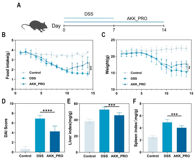
Zhang et al. (2025), original Figure 1, reproduced in full under CC BY 4.0. Twenty-four male C57BL/6J mice were allocated to Control, DSS and AKK_PRO groups, eight mice per group, for 14 days. DSS-induced injury occurred during the first seven days and intervention covered the full period. A: timeline; B: food intake (g); C: body weight (g); D: disease activity index (DAI); E/F: liver and spleen indices (mg/g). Means ± SD; n = 8 mice per group. **p < 0.01, ***p < 0.001 and ****p < 0.0001 apply to the bracketed comparisons in the original figure. Because intervention included the induction period, this is not a treatment started only after disease was established. [4]
The three-group profiles and DAI show the model injury and a different response in the intervention group. Not every AKK_PRO endpoint returns to the control level. The result concerns a defined inactivated strain in chemical colitis, rather than an established human treatment effect.
Immune and microbiota findings depend on context
Immune modulation does not mean suppressing every cytokine or increasing every immune response. Cellular constituents, host status and the inflammatory stimulus can alter the direction of a measured response. In colitis models, reduced inflammation often accompanies preservation of the mucosa. Microbiota and fecal short-chain fatty acid measurements in the AKK PROBIO study can be read alongside tissue findings, but correlations and predicted functions do not establish that a particular genus or pathway is a necessary mediator. [1, 4]
For this reason, increasing beneficial bacteria is not a sufficiently precise mechanistic description. A higher relative abundance does not necessarily mean a higher absolute cell count, and predicted metabolic functions are not measured metabolic fluxes. A more informative question is which preparation changes which measurable physiological outcome in which host context. [2, 4]
Human outcomes depend on the preparation and endpoint
In an eight-week randomized, double-blind trial, Andresen and colleagues assigned 443 adults with irritable bowel syndrome to heat-inactivated Bifidobacterium bifidum MIMBb75 or placebo. The composite primary endpoint required at least 30% improvement in abdominal pain together with adequate relief of overall symptoms in at least four of eight treatment weeks. It was reached by 74/221 participants versus 43/222, reported as rounded rates of 34% and 19%. The risk ratio was 1.7 (95% CI 1.3–2.4; p = 0.0007). This supports a symptom benefit for the specified inactivated preparation, rather than a shared therapeutic effect of all postbiotics. [6]
Metabolic findings also depend on the population and endpoint. A 2026 trial in 90 adults with overweight or obesity began with an eight-week low-energy diet and at least 8% weight loss, followed by a 24-week maintenance period. Weight regain was 1.2 kg with pasteurized A. muciniphila MucT and 3.2 kg with placebo (p = 0.012). The result concerns maintenance after diet-induced weight loss; it is not a direct estimate of weight loss without the preceding dietary intervention. [7]
A separate multicenter trial randomized 143 participants, of whom 142 received at least one administration of study product. After four months, the primary endpoint, whole-body insulin sensitivity assessed by the Matsuda index, did not differ significantly between groups. Signals in subgroups such as participants with low baseline Akkermansia levels were subsequent exploratory findings and cannot replace the primary result. A reported body-weight p value of 0.06 should not be described as a significant reduction. These trials address different questions and must retain their respective conditions. [8]
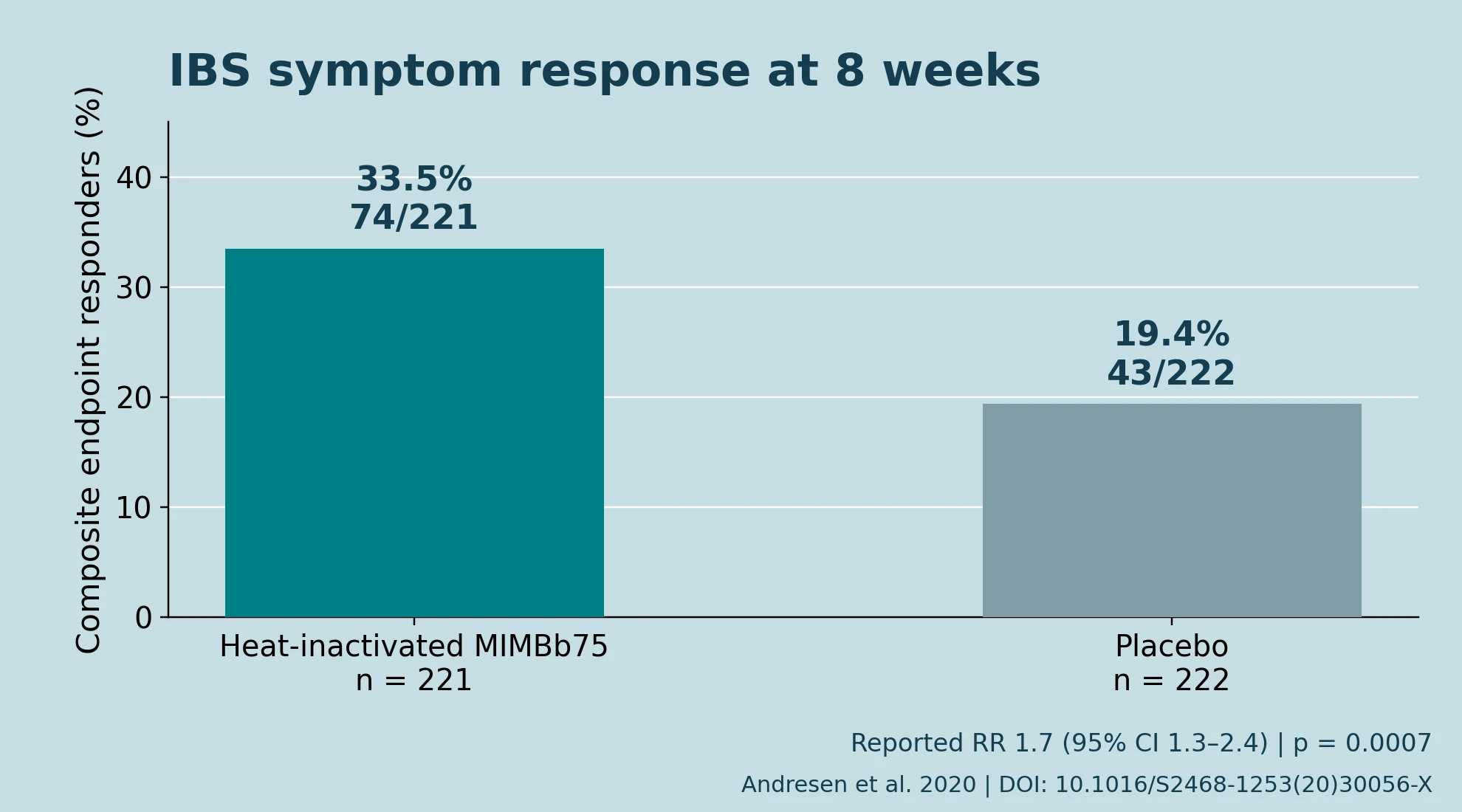
Independently plotted from participant counts in the primary abstract of Andresen et al. (2020): HI-MIMBb75, 74/221; placebo, 43/222. Adults with irritable bowel syndrome were assessed over eight weeks. Calculated proportions are 33.5% and 19.4%; the paper reports rounded values of 34% and 19%. The composite endpoint required at least 30% improvement in abdominal pain and adequate relief of overall symptoms in at least four of eight weeks. The reported risk ratio was 1.7, 95% CI 1.3–2.4; p = 0.0007. Bars show proportions without SD or SEM error bars; individual-level data were not available. This factual redraw does not reproduce publisher artwork. [6]
Compare response proportions using each complete group denominator. The difference is approximately 14.1 percentage points, calculated from the counts. Most participants in the intervention group did not meet the composite endpoint; the result is not a universal cure or a comparison with live bacteria.
Processing defines material identity and application value
Inactivation is more than reducing a viable count to zero. The strain, fermentation matrix, inactivation process and drying method jointly shape the cellular structures and soluble constituents in the final preparation. Materials made from the same named species may have different functions. Changing the matrix, process or strain of a clinically studied preparation requires a renewed connection between material characteristics and evidence of benefit. [1, 2, 5]
Quality assessment can be organized into three levels: establish strain and matrix provenance; verify inactivation status and compositional consistency using suitable methods; and determine whether health benefits are supported for the same preparation, relevant population and intended context. Inactivation may reduce the burden of maintaining cell viability, but it does not guarantee room-temperature stability, processing tolerance or a long shelf life. These are questions for product specifications and stability data. [1, 2]
The absence of live cells also does not establish suitability for every population. The source organism, cellular constituents, fermentation matrix and process residues remain part of safety assessment. Good short-term tolerability cannot substitute for long-term or population-specific data. The practical value of postbiotics lies in connecting an identifiable non-viable microbial preparation with reproducible quality and a measured host benefit, so that each claim has a defined scope. [1, 2, 6]
Table 2. What the endpoints measure
| Measurement | Supported interpretation | Interpretive boundary |
|---|---|---|
| Protein or gene expression | Structural and regulatory clues | Does not replace permeability testing [3, 5] |
| TEER or tracer flux | Barrier function in a cell-layer model | Not a clinical or human absorption outcome [3, 5] |
| Mouse DSS pathology or DAI | Tissue and disease findings in chemical colitis | Retain strain and model context [4] |
| Human symptom or metabolic primary endpoint | Clinical result for a defined preparation and population | Exploratory subgroups do not replace the primary endpoint [6, 7, 8] |
References
[1] Salminen S, Collado MC, Endo A, Hill C, Lebeer S, Quigley EMM, Sanders ME, Shamir R, Swann JR, Szajewska H, Vinderola G (2021). The International Scientific Association of Probiotics and Prebiotics (ISAPP) consensus statement on the definition and scope of postbiotics. Nat Rev Gastroenterol Hepatol 18(9):649-667. doi:10.1038/s41575-021-00440-6 Corrections: 10.1038/s41575-021-00481-x; 10.1038/s41575-021-00506-5; 10.1038/s41575-022-00628-4
[2] Vinderola G, Sanders ME, Cunningham M, Hill C (2024). Frequently asked questions about the ISAPP postbiotic definition. Front Microbiol 14:1324565. doi:10.3389/fmicb.2023.1324565
[3] Gao J, Li Y, Wan Y, Hu T, Liu L, Yang S, Gong Z, Zeng Q, Wei Y, Yang W, Zeng Z, He X, Huang SH, Cao H (2019). A Novel Postbiotic From Lactobacillus rhamnosus GG With a Beneficial Effect on Intestinal Barrier Function. Front Microbiol 10:477. doi:10.3389/fmicb.2019.00477
[4] Zhang H, Liu C, Huang Y, Ma X, Ren D (2025). Inactivated Akkermansia muciniphila AKK PROBIO Preserves Intestinal Homeostasis and Ameliorates DSS-Induced Colitis in Mice. Foods 14(23):4063. doi:10.3390/foods14234063
[5] Guo J, Zhao Y, Guo W, Sun Y, Zhang W, Zhao Q, Zhang Y, Jiang Y (2025). Effects of Lactobacillus paracei JY062 Postbiotic on Intestinal Barrier, Immunity, and Gut Microbiota. Nutrients 17(7):1272. doi:10.3390/nu17071272
[6] Andresen V, Gschossmann J, Layer P (2020). Heat-inactivated Bifidobacterium bifidum MIMBb75 (SYN-HI-001) in the treatment of irritable bowel syndrome: a multicentre, randomised, double-blind, placebo-controlled clinical trial. Lancet Gastroenterol Hepatol 5(7):658-666. doi:10.1016/s2468-1253(20)30056-x
[7] Mount S, Canfora EE, Jocken JW, Umanets A, Hul G, Coenjaerds M, Aldaz Laquidain P, Adriaens ME, Holst JJ, Jardon KM, Segers A, Suenaert P, de Vos WM, Blaak EE (2026). Pasteurized Akkermansia muciniphila MucT for weight loss maintenance in people with overweight and obesity: a controlled randomized trial. Nat Med 32(6):2107-2116. doi:10.1038/s41591-026-04394-7
[8] Suenaert P, Segers A, Rymenans L, Devroye H, Moll JM, Cani PD, de Vos WM (2026). Effect of pasteurized Akkermansia muciniphila MucT on insulin sensitivity, body composition, and GLP-1 production in subjects with metabolic syndrome: impact of low baseline gut Akkermansia levels. Gut Microbes 18(1):2690689. doi:10.1080/19490976.2026.2690689